In 1999, Dr Malgorzata Lekka presented the first AFM results that proved cancerous cells have different mechanical properties to normal cells.
That she did this working within an institute that specialised in Nuclear Physics rather than biology and on a ‘home-built’ Atomic Force Microscope that had to be modified makes this achievement all the more outstanding.
Dr Lekka, now professor and department head at the Henryk Niewodniczański Institute of Nuclear Physics, Polish Academy of Sciences in Kraków (Poland), started working with AFM almost 30 years ago this October. It was down to her scientific mentor and supervisor, Dr Zbigniew Stachura, who decided: ‘You have a medical physics background, so let’s start with the AFM to look at cancer cells’. This was the moment when her long-standing collaboration with Prof. Piotr Laidler's group from Collegium Medicum of the Jagiellonian University started. Initially, they provided cancer cells and expertise in cell biology, including how to culture them. As Lekka explains, in reality, she had a degree in Physics with a specialisation in Medical Physics, having taken some associated courses in radiology and similar. Notwithstanding, she began tackling the project head on.
Home-built construction
“There were only a few AFMs in Poland at that time, but the one I began working with was a home-built construction designed for another project. The first task was to redesign the mechanical construction from vertical to horizontal to be able to work with liquid samples. It was a 90-degree conversion – easy to say, hard to do!” says Lekka.
“In the beginning, it was not about looking at cell mechanics, I was just imaging, which seems silly looking back now, especially with our home-built AFM. The resolution of the AFM wasn’t sufficient to obtain nice images of the cell membrane. Now we know that cell membranes can be seen using High-Speed AFM – but our AFM definitely wasn’t high speed back then.”
Whilst Lekka was working in a post-Communist political environment in Poland, as she explains at the time, getting papers and journals was difficult because of the lack of money to pay for subscriptions. Additionally, no one specialised in AFM and biology in Poland back then. Lekka’s route to gaining knowledge in the area was as homebuilt as the AFM she used.
“It was kind of difficult,” says Lekka, “because in Poland, most AFMs were used for surface physics. There was no biological AFM. My mentor talked to other colleagues, including Prof. Günter Schatz from the University of Konstanz in Germany, who invited me to go there for a few 10-day visits to use their library. This 10 days in the library there to read up on the most recent papers on what was going on with AFM and biology was very helpful for me.”
Home-build AFM
Cell Mechanical Properties
As Lekka was coming to the end of her PhD it became increasingly apparent imaging wasn’t working. That was when she started to think about looking at the mechanical properties instead.
“This was a kind of ‘Bingo’ moment,” says Lekka, “I began measuring the mechanical properties of the living cells and quickly discovered I was able to generate interesting results, showing that cancer cells are softer than normal cells.”
Those results and the paper that derived from them were first published in Electron Technology in 1997, a Polish journal. By 1999 it was published in the European Biophysics Journal, and her work gained international attention.
AFM and Muscular Dystrophy
After her PhD, Lekka continued to work with AFM, looking further into its uses from a biological perspective. From the earliest days, she was keen to understand ‘how can we use this technique for diagnostic purposes?’
Her work on tissue samples – which split her time between IFJ PAN and École Polytechnique Fédérale de Lausanne (EPFL, Switzerland) - was a major study looking at Muscular Dystrophy in mice, being an interest of Prof. Nicolas Mermod's group. Thanks to Dr Andrzej Kulik (EPFL), it was possible to obtain financing from both EPFL and NATO, which gave Lekka better access to human and animal samples in her work.
“Working with human and animal samples is difficult, you can only do it with other people who have the skills and also the permission to work with human and animal tissue,” says Lekka.
“Working on Muscular Dystrophy allowed me to master my abilities. I had real tissue to measure, great access to samples, and it was nice to work and to talk with other groups that use AFM, especially from Prof. Giovanni Dietler's group.”
Principles of AFM measurements of muscle resistance to deformation. Taken from https://www.cell.com/molecular-therapy-family/molecular-therapy/fulltext/S1525-0016(16)31470-8
Much has changed in the past thirty years. Lekka’s work has been well-respected and supported within the IFJ PAN, leading to the development of her own lab and then her own department. A collaboration with nearby scientific groups enabled Lekka's group to work with animal and human materials, thus enabling Lekka and her team to have greater control over the samples they are working with, a subject Lekka feels strongly about:
“I don’t want the PhD students to measure only samples from collaborators because sometimes samples are good for AFM measurements, sometimes not, and there might not be enough time to complete a thesis within 4 years. That is why we started to culture cells in my group and combine these two classes of samples.”
AFM Protocol Development
The aim of the most recent projects in which Lekka's group participated was the EU-funded Phys2BioMed Marie Curie ITN network coordinated by Prof. Alessandro Podestà from Milano University in Italy , which offered interdisciplinary and cross-sectoral training to a group of motivated early-stage researchers (ESRs) on the application of mechanics cells and tissues in clinical samples, aiming at developing novel early-diagnostic tools.
Within this project, one of the aims was to develop a protocol and criteria that allow AFM to be used in clinical settings, as reported in the prepared and submitted manuscript (https://www.biorxiv.org/content/10.1101/2023.06.14.544753v1), showing that within the standardized protocol of elasticity measurements - starting from sample preparation and measurements data analysis – it is possible to get similar results regardless which laboratory is involved.
“In fact, this work started in 2010 with the COST Action TD1002. The one aim of this network was the standardization of the AFM calibration, which resulted in the SNAP procedure (https://www.nature.com/articles/s41598-017-05383-0). The work on standardization realized within Phys2BioMed goes further. We identified several factors that affect the outcome of the mechanical measurements,” says Lekka, “including sample preparation, data analysis, etc.” The Phys2Biomed project enables me to join the editors of the book "Mechanics of Diseases: Biomedical Aspects of the Mechanical Properties of Cells and Tissues", i.e., renowned AFM scientists Prof. Daniel Navajas (University of Barcelona), Manfred Radmacher (Bremen University) and Alessandro Podesta (University of Milano).
Comparison of mechanical properties of polyacrylamide gels measured with conventional procedure (A) and with the standardized nanomechanical AFM procedure SNAP (B). Taken from https://www.nature.com/articles/s41598-017-05383-0
Mimicking the Tumour Environment
Dr. Lekka’s latest research efforts are on trying to mimic the tumour environment within the project financed by National Science Center (NCN, Poland). “We’re starting to form and modify spheroids,” says Lekka, “we’re making the combination of normal cells, or reference cells plus cancerous ones, because when we look at cancer progression, this is a very inefficient process. There’s a lot of cancerous cells which are in our organisms but not all of them are producing metastasis. So then there is the question, what are the conditions or criteria that this singular cell or particular group of cells form metastasis?”
Lekka is keen to model the environment, embed the spheroid inside the hydrogels, and see what happens.
“My hypothesis is it is not only the chemistry or biomechanics of molecular or genetic changes involved in forming metastasis. We are moving, so we are producing compressive tension and shear forces continuously. I hypothesize that a very low shear force triggers the cells forming metastasis.”
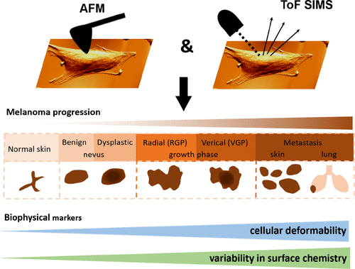
Even in her latest research, Dr. Lekka continues to look for new ways of integrating other techniques with AFM, like, for instance, time of flight secondary ion mass spectroscopy (ToF SIMS, https://pubs.acs.org/doi/10.1021/acs.analchem.9b01542), constantly finding ways of pushing the boundaries of what is currently possible to further our exploration of the unknown in the arena of cancer. To obtain a full image of cancer progression, she is collaborating with Prof. Katarzyna Rolle from the Department of Molecular Neurooncology at the Institute of Bioorganic Chemistry PAN in Poznan to try to correlate the changes in spheroid mechanics with alterations in gene expression.
Studying the relation between the surface and biomechanical properties of melanoma cells, measured by mass spectrometry (ToF-SIMS) and atomic force microscopy (AFM). Taken from https://pubs.acs.org/doi/10.1021/acs.analchem.9b01542
Her focus remains on the diagnostic possibilities this research could lead to by combining and developing techniques to transform what currently would be a ‘needle in a haystack’ exercise into a means of being able to determine whether a person is likely to form metastasis or not.
Women in AFM
Dr Lekka’s experience of being a woman in a domain where AFM is used is a fantastic one. “For many years, my whole team is just women!” she says, “or rather, it used to be. We have two male members in my group right now, but we were only women for many years. The group is small, maximum of 10 people, depending on the number of PhD students. I think that biological applications of AFM are attractive for all researchers who like cooking – that’s not to lower the significance of AFM by saying this, though. You have to prepare the samples in a special way. You can go through the protocols (that are like cooking recipes), but this is not interesting, but if you have to change something, this is fun. Regarding the measurements, there is, of course, the protocol to follow at the beginning, but then you can try to do something different. That’s why it’s kind of cooking, there’s a recipe, but everyone does it their own way.”
Lekka’s approach allows her student to do almost everything, from sample preparation to data analysis and interpretation. There are weekly discussions to plan the work for the week ahead, but then they have the opportunity to be self-directed.
“We can work end-to-end, from cell culture to analysis of samples, to experience a real correlation between physics and biology. Our main method of work is AFM, but we also use fluorescent microscopy with live cell imaging capability that allows us to study, e.g., cell migration. We are using a rheometer for macro-mechanics. We can perform all biological assays using an ELISA reader or Western blot. The majority of PhD students that were and are in my group are not physicists – we have the physics method, and I’m a physicist, but my students are mostly biology-related,” says Lekka.
Lekka also professes a very supportive style of working within her department.
“At the beginning, I try to ensure that my students have initial successful experiments to encourage them to use AFM and think about what they can do further,” says Lekka.
“I have two AFMs. One is very straightforward and relatively simple to use. Once they know how to use it, they switch to a more sophisticated machine, a real scientific machine where you have to know what you are doing. The software is very well developed. There are a lot of possibilities to measure, and you have to know what you are doing.”
Conclusion
Whilst it might have been curiosity that initially drew Lekka to working with AFM, it was the enjoyment of the intellectual challenge that kept her there – and of course, the knowledge that working with disease had the potential to offer practical and much-needed real-world solutions.
“Once you go deeper into technique, you find the answers you have in your mind are not quite right. Then you have fun finding out why your thinking is wrong! To this day, I am always curious why the AFM measurements don’t match my expectations – and always, there are some really surprising findings.”
“I’m a very pragmatic person. I wouldn’t be a good theoretical physicist because the outcome doesn’t have a practical application. Theory is important however,” says Lekka.
“Most of my PhD students had to pass the exam for Quantum mechanics – they say that it is not needed, but this is not true. You need to train the brain to think in a very abstractive way, and quantum mechanics is great for this.”
Lekka’s greatest wish is to one day see her technique in use in a hospital. “That would be perfect!” she says.
This wish isn’t necessarily a far-fetched one. There are some trials currently taking place for the development of a clinical diagnostic tool. “Maybe one day before I die, I will see AFM in use in the hospital,” says Lekka, “I’d certainly be the first to pay for such an examination.”
Dr Lekka’s department website: https://www.ifj.edu.pl/oddzialy/no5/nz55/en/home/
Twitter: @lekka_lab
If you enjoyed this blog post, you might also like
NuNano Interviews: Liisa Lutter, Postdoctoral Researcher at UCLA (Women in STEM series),


