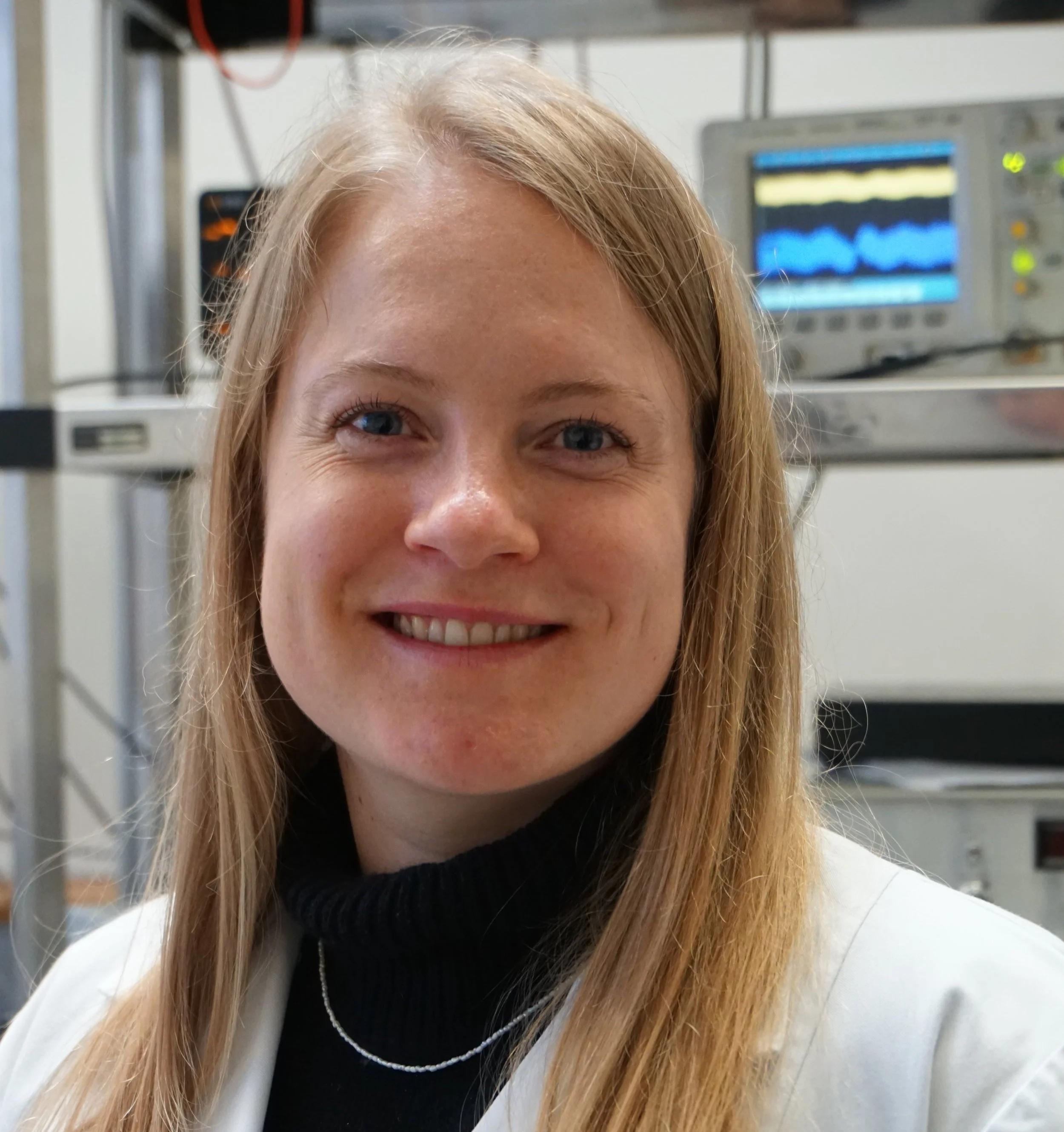
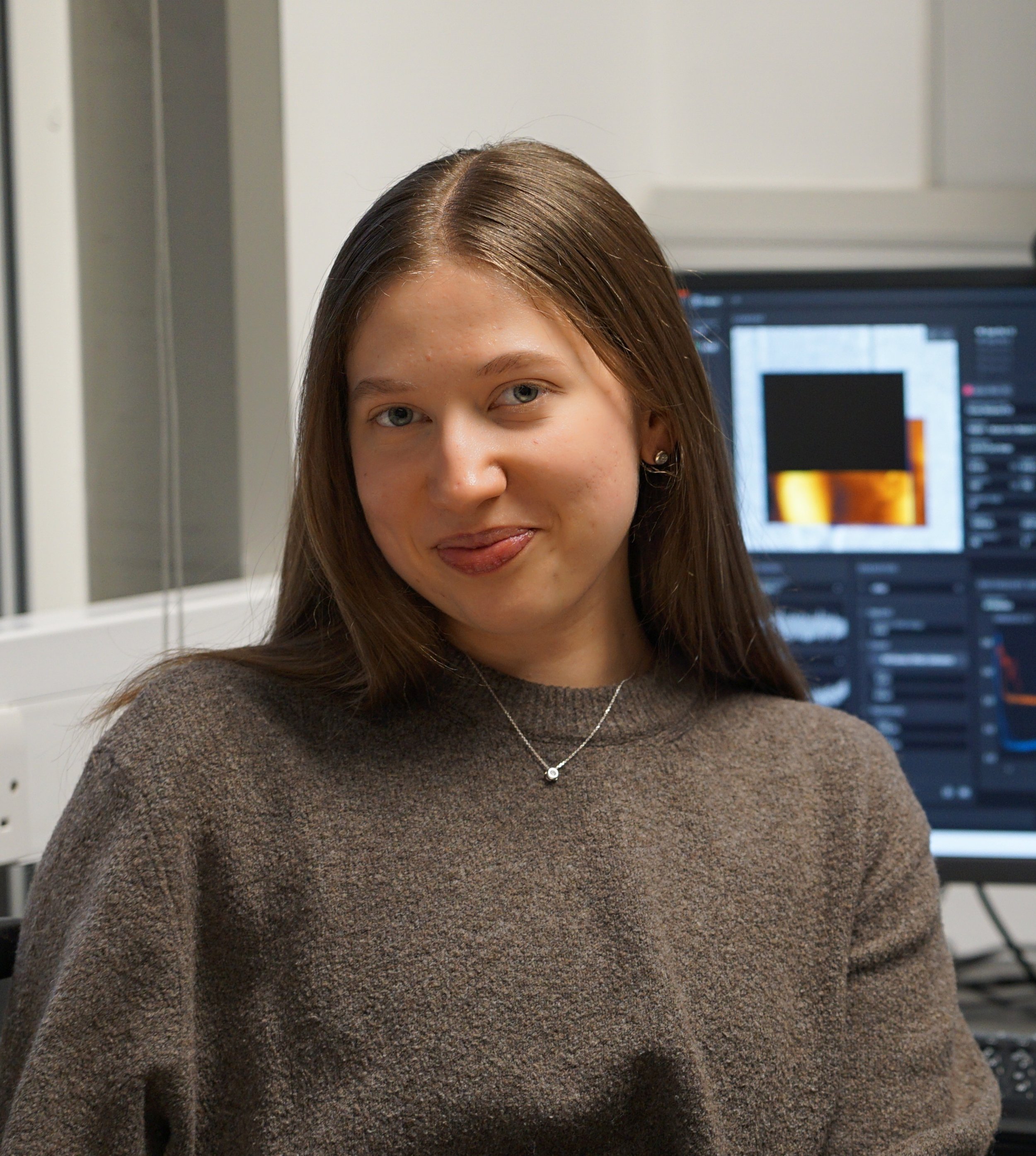
We are thrilled to have previous ‘NuNano Woman in AFM’ Dr Lekshmi Kailas introduce the 2025 list of brilliant female scientists you need to know about.
I am delighted to introduce the NuNano 2025 Women in AFM celebration because I believe platforms highlighting women’s achievements are important in boosting confidence in the younger generation as they enter STEM education. There are fewer women in STEM, not because these fields are difficult for women, but I believe because of misconceptions at a very early stage that dissuade girls and young women from taking up these subjects. Platforms like this, which put the spotlight on women already in STEM, can help counter such outdated narratives.
Whilst it is true that many women experience negative bias within science and higher education, I feel very fortunate that this has not been my experience so far. As an Indian woman living and working in a different country, my story could’ve been very different. However, my positive experience only makes me champion the cause of women even more. It pains me to hear about the different types of discriminatory behaviour that people, especially women, experience. Because I didn’t experience any discrimination based on my gender or race in higher education the way many others have, it seems self-evident to me that no one should have to.
Formative Support
Education has been one of the biggest priorities in my family. My parents made sure that my brother and I got the best education possible and that has immensely helped us pursue the careers of our choice. In my maternal family, my grandfather and my great-grandfather were all school headmasters. My grandfather was the one who first piqued my interest in science. He was an advocate of learning science and physics in particular. He used to say if you understand physics, it makes it easier to understand other sciences. I think that made me gravitate towards science in general and physics in particular.
Even in school, in college and in university back in Kerala, my home state in India, I never felt discriminated because of my gender. In fact, there were more girls than boys in my B.Sc. Physics, M.Sc. Physics and B.Ed in Science Education classes and the teachers treated us all equally. I firmly believe that the level of support received at that formative stage from my family and my teachers helped me in making my career choice to stay on in science.
For my career journey into scientific research, I would like to thank Prof. Laly Pothen who was my college Chemistry professor as well as a very close family friend. Initially, I was doubtful whether I'd be a good fit in research, but being a science researcher herself, Aunt Laly reassured me that I would be very suited to this environment. She and Prof. Sabu Thomas of Mahatma Gandhi University have been my rocks of support throughout my career journey so far.
Introduction to AFM
I did my PhD at Université Catholique de louvain in Belgium in the group of Prof. Patrick Bertrand, which is when I first came across an AFM. I certainly wouldn’t say I loved working with AFM from the very beginning, but I didn't dislike it either. It was a technique I needed for my studies, and I didn't think too much about it. AFM became the main technique when I joined the group of Prof. Jamie Hobbs in Sheffield for my first post-doc to look at polymer crystallization and nucleation behaviour.
It was there I discovered that once you get more proficient with it, AFM becomes this amazing window into the nanoworld. You can view the surfaces at high resolution, check the forces acting at the nanoscale and even get mechanical properties of tiny areas; you can work in liquid, or air, without the need for vacuum or any special sample preparation and you get a 3D image after a single scan. Even after I moved to Prof. Per Bullough’s group in Sheffield to use CryoEM to look at the sub-nanometer architecture of exosporium layers, I found myself going back to AFM to get information to complement the EM data.
During my time as a visiting scientist with the Department of Atomic Energy, Govt. of India at IGCAR and as an instrument scientist at the Bernal Institute of the University of Limerick in Ireland, I concentrated primarily on AFM.
Gifted researchers & enthusiastic students
Currently, I am the Experimental officer in AFM at the School of Physics and Astronomy of the University of Leeds, and I manage the AFM Facility at the Bragg Centre for Materials Research. The AFMs here are used by students and researchers coming from different Schools including Physics, Chemistry, Engineering, Medicine, Biology, Food Science, and even Design. I am happy to say that lots of female researchers come to use our Facility and the gender ratio is not so lopsided. It is wonderful to interact with gifted researchers and enthusiastic students from different disciplines, irrespective of any consideration to their gender. At work, I am surrounded by people who share my interest in science and so, I really enjoy working in this field and am happy tAo do it long term.
Positive Environment
It is my firm belief that irrespective of gender or race or any other consideration, young people should get into STEM if they have a passion for these fields. My experiences have taught me that you never know what you are capable of until you try. If you find that something is difficult at first, don’t give up. Try again. Failure is also part of the learning process, and it helps to look at the problem from a different perspective and sometimes come up with an even better solution.
I recognise there are lots of people around me who have made me who and what I am now. I believe that I was able to do this journey because of the positive environment I was exposed to throughout. I really hope that young people, especially young women, who are going to come into this field will also experience the same. It is on each of us to behave in respectful and supportive ways with one another without any bias. In this way, we create the world we want to live and work in and make the environment better for everyone – which in turn will benefit the broader aims and achievements of the science we are exploring.
So, come along. Let us go and take a look at some of the amazing women scientists, their journeys and the wonderful work they have been doing, and let us get inspired.
Click the images below to read about each researcher’s work and their career.
Latest AFM Papers
If you know of any women, or other marginalised and underrepresented people that are doing amazing work with AFM, who might be interested in being included in our future profiles please do get in touch with us community@nunano.com


