Women in AFM

Sarah Stainer

Sarah Stainer is a PhD student at the Johannes Kepler University Linz (JKU) and is currently finishing her thesis on real-time visualization of highly mobile proteins using high-speed AFM (HS-AFM). During her PhD, she specialized in the imaging of highly mobile proteins, studying their flexibility and conformational changes. She has always been fascinated by the complex workings of cells and wanted to understand their mechanisms at the molecular level.  HS-AFM fascinates her because you can observe very small biological samples such as proteins and DNA and watch their movement in real time. Resolving the features of flexible domains can be challenging using conventional structural determination techniques. HS-AFM can add new information about dynamics and flexibility to protein structures that would otherwise be described by a static representation.


Sarah Stainer


Recent AFM-related papers:

Biography: Sarah grew up in Salzburg, Austria and studied Molecular Biology in a joint program at the Paris-Lodron University of Salzburg and the Johannes Kepler University of Linz. She was introduced to AFM during her master thesis at the Johannes Kepler University, where she studied the surface pattern of rhesus factor membrane proteins on erythrocyte cells using Topography and Recognition Imaging (TREC). During her master thesis, she applied for a PhD position at JKU and joined the Applied Experimental Biophysics group, specializing in Single Molecule Force Spectroscopy (SMFS), Topography and Recognition Imaging (TREC) and High-Speed AFM (HS-AFM). She spent half a year abroad in Japan as a research visitor to deepen her knowledge in HS-AFM.

LinkedIn: www.linkedin.com/in/sarah-stainer-b1473b142

Webpage: https://www.researchgate.net/profile/Sarah-Stainer


Are you a woman conducting AFM research or know of someone you would like to nominate to be featured in our next #WomenInAFM campaign? Contact us at community@nunano.com!

Prof Kerstin Blank

Kerstin Blank is a Professor at the Institute of Experimental Physics at Johannes Kepler University Linz, Austria, where she leads the Division of Biomolecular and Selforganizing Matter. Originally trained as a protein biochemist, she was introduced to atomic force microscopy (AFM) during her PhD in Hermann Gaub’s lab at Ludwig Maximilian University Munich. Since then, she has been fascinated by the intricate relationship between protein structure and mechanics.

Her current research focuses on the mechanical properties of structural proteins that form biological materials. Using these proteins as blueprints, she designs mechanoresponsive nanomechanical building blocks. A major focus is the de novo engineering of coiled coils and their mechanical characterization via single-molecule force spectroscopy. Integrating additional functionalities, such as optical readouts of their molecular state, she employs these coiled coils as molecular force sensors and as mechanoresponsive elements in smart hydrogels. These tools provide new insights into mechanical signaling at cell-material interfaces, both in 2D and 3D cell culture environments.


Kerstin Blank - Photo Credit: Sabine Starmayr


Recent AFM-related publications:

Biography: Kerstin Blank received her PhD in Biophysics from Ludwig Maximilian University Munich in 2006. After brief postdoctoral stays in Strasbourg and Leuven, she became an assistant professor at Radboud University Nijmegen in 2009. In 2014, she joined the Max Planck Institute of Colloids and Interfaces in Potsdam, where she led the Max Planck Research Group Mechano(bio)chemistry. Since October 2021, she has been a full professor at Johannes Kepler University. Kerstin Blank co-founded and chaired the Gordon Research Conference on Multiscale Mechanochemistry & Mechanobiology. Since 2022, she has served as (deputy) speaker of the Biological Physics section of the German Physical Society. She is also a member of the editorial board of RSC Mechanochemistry.

Website: www.jku.at/biom

LinkedIn: https://at.linkedin.com/in/kerstin-blank-b294269


Are you a woman conducting AFM research or know of someone you would like to nominate to be featured in our next #WomenInAFM campaign? Contact us at community@nunano.com!

Alyona (Olena) Yedelkina

Alyona (Olena) Yedelkina is a Ph.D. student in Dr. Antonio Benedetto’s NanoBioPhysics lab at University College Dublin, Ireland. Her research explores the interactions between ionic liquids (ILs) and cell membranes using atomic force microscopy (AFM), neutron scattering, and other biophysical techniques.

Working with synthetic lipid bilayers of varying complexity, she investigates how ILs influence membrane properties. Through AFM, she examines structural changes and mechanical responses to better understand IL effects at the nanoscale. Collaborating with her research group, she aims to correlate IL-induced changes in cell metabolism and mechanics with the mechanical and structural data obtained from membranes. Alyona hopes her research will lead to a better understanding of cell membrane biophysics and pave the way for IL applications in nanotechnology and medicine.


Alyona (Olena) Yedelkina


Biography: Alyona’s journey into biophysics began in 2019 in Slovakia, where she earned a degree in Biophysics at Pavol Jozef Šafárik University and conducted research on protein molecular properties and interactions at the Center for Interdisciplinary Biosciences in Košice. During this time, she gained experience with a variety of biological assays and biophysical techniques, studying enzyme activity, stability, and aggregation under different physical conditions. In 2022, Alyona moved to Dublin to join Dr. Antonio Benedetto’s research group at University College Dublin. Since starting her Ph.D., her focus has shifted to the biophysics of cellular membranes, with atomic force microscopy (AFM) becoming her primary technique. She now uses AFM, along with neutron scattering and other biophysical approaches, to investigate the interactions between ionic liquids and biological membranes.


X: @AYedelkina

LinkedIn: https://www.linkedin.com/in/alyona-yedelkina-147b92328/

Website: https://www.antoniobenedetto.eu/Members/


Are you a woman conducting AFM research or know of someone you would like to nominate to be featured in our next #WomenInAFM campaign? Contact us at community@nunano.com!
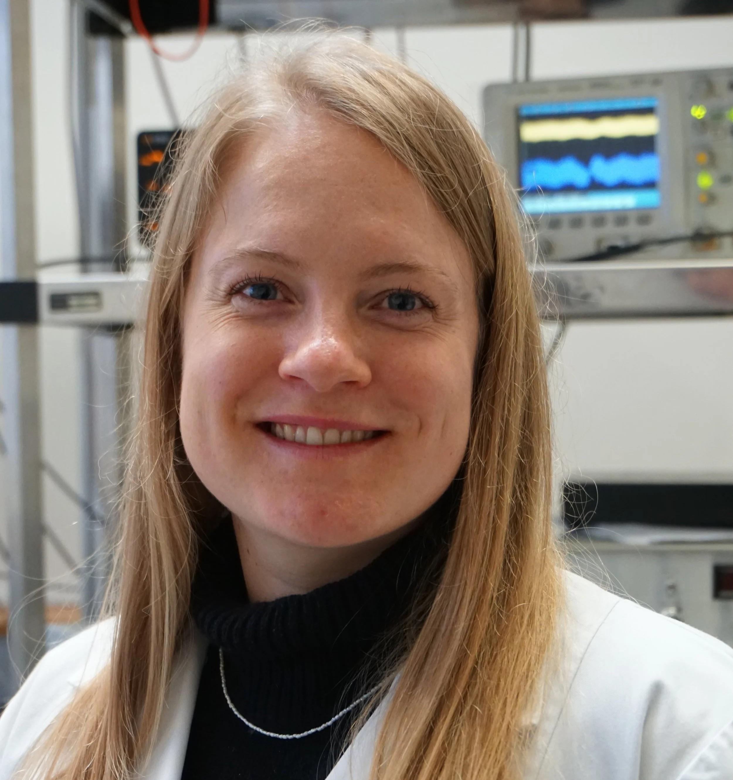
Dr Lorena Redondo Morata

Lorena Redondo Morata is a researcher at the DyNaMo U1325 laboratory at Aix-Marseille Université & INSERM in Marseille, France. Her research focuses on understanding membrane remodeling processes and how proteins and lipids drive membrane dynamics, including deformation, fusion, and fission. These biological processes often occur on timescales ranging from sub-seconds to minutes.

What excites Lorena most about atomic force microscopy (AFM) is its unique ability to explore structure, mechanics, and dynamics in a controlled environment without the need for labeling. Given the inherent complexity of cell membranes, her research strategy centers on developing model membrane systems that mimic cellular membranes with controllable complexity.

Currently, Lorena's research is dedicated to investigating the mechanics and dynamics of cell membrane remodeling using AFM and correlative microscopy techniques. Her key research interests include studying the nanomechanics of membrane disruptors, such as antimicrobial peptides and surfactants, as well as exploring extracellular vesicles and the dynamics of lipid nanodomains, particularly in the context of membrane fusion and remodeling processes.


Lorena Redond Morata


Recent papers:

Biography: Lorena obtained her PhD in Physical Chemistry from the University of Barcelona, Spain, in collaboration with the Institute for Bioengineering of Catalonia, focusing on lipid bilayer nanomechanics using AFM-based force spectroscopy. After earning her PhD in 2012 under the supervision of Fausto Sanz, she joined Simon Scheuring’s lab at Inserm in Marseille to study biomembrane dynamics with High-Speed AFM. Awarded the Young Researcher Prize by the Spanish Biophysical Society in 2016, she became an Inserm researcher at the Institut Pasteur in Lille in 2018. In 2024, she relocated her laboratory to the DyNaMo Inserm unit in Marseille, where she focuses her research on lipid membrane nanomechanics and the dynamics of lipid nanodomains in the context of membrane fusion or other remodeling processes.

BlueSky: @lorenaredondo.bsky.social

LinkedIn: https://www.linkedin.com/in/lorena-redondo-morata-06a89051/

Website: https://sites.google.com/view/fm4b-lab/home?authuser=0


Are you a woman conducting AFM research or know of someone you would like to nominate to be featured in our next #WomenInAFM campaign? Contact us at community@nunano.com!

Dr Carmen Munuera

Carmen Munuera is a Tenured Scientist at the Materials Science Institute of Madrid, ICMM-CSIC, Spain. She is the head of the Scanning Probe Microscopy laboratory of the 2D Foundry group and is expert on the advanced modes of the technique, such as Kelvin Probe, Piezo Force and Magnetic Force Microscopy. Her research focuses on the characterization and manipulation of functional properties across diverse systems, with a particular emphasis on nanostructures and interfaces. The systems investigated during her research trajectory range from self-assembled monolayers (SAMs) and molecular switches to complex oxide heterostructures and superconducting materials. Presently, her primary research focus revolves around the exploration of 2D materials and heterostructures. Utilizing Scanning Probe Microscopy, she conducts in-situ characterizations of these systems and devices under challenging conditions, such as in-operando or under strain, scrutinizing the induced effects at local scale.


Carmen Munuera


Recent AFM-related papers:

Biography: Carmen completed her undergraduate studies in Physics at the University of Seville, Spain, earning her Bachelor's degree, along with the Extraordinary Bachelor's Award from the Faculty of Physics. She pursued her doctoral training at the Institute of Materials Science in Madrid (ICMM-CSIC), obtaining in 2007 the PhD title from the Autonomous University of Madrid. Her doctoral research, supervised by Prof. Carmen Ocal,  focused on the relationship between functional and structural properties in self-assembled monolayers, utilizing the Scanning Probe Microscopy technique.

She carried out a first postdoctoral stay at the Material Science Institute in Barcelona (ICMAB-CSIC) where she helped Prof. Ocal setting up the SPM laboratory of the current “Group of Physical Chemistry of Surfaces and Interfaces”. In 2008 she became a postdoctoral fellow at Max-Planck Institute for Metal Research, in Stuttgart. Her research interests reoriented after this postdoctoral stage, focusing on the nanoscale study of complex oxides heterostructures and the novel properties arising at the interfaces in these systems.

In 2010 she moved back to the ICMM joining the group of Prof. Mar García-Hernández, to set up a three axis vector magnet system for cryogenic scanning probe microscopy. This system was crucial to her investigation and motivated her interest in one of her actual research lines: the characterization and manipulation by MFM of the vortex lattice in layered superconductors.

Since 2017, she has been part of the 2DFoundry group at ICMM, heading the Scanning Probe Microscopy laboratory and focusing on the study of van der Waals materials.

Carmen's research contributions have been consistently recognized, with fellowships obtained through open and competitive calls supporting her various research stages (FPU, Postdoctoral MEC, Juan de la Cierva and Ramón y Cajal)  In 2011, she was honored with the Young Scientist Award from the Royal Science Academy of Seville for her outstanding research work.

Twitter: @CarmenMunuera1

LinkedIn: https://www.linkedin.com/in/carmen-munuera-lopez-306b27200/

Webpage: https://sites.google.com/view/munueralab


Are you a woman conducting AFM research or know of someone you would like to nominate to be featured in our next #WomenInAFM campaign? Contact us at community@nunano.com!
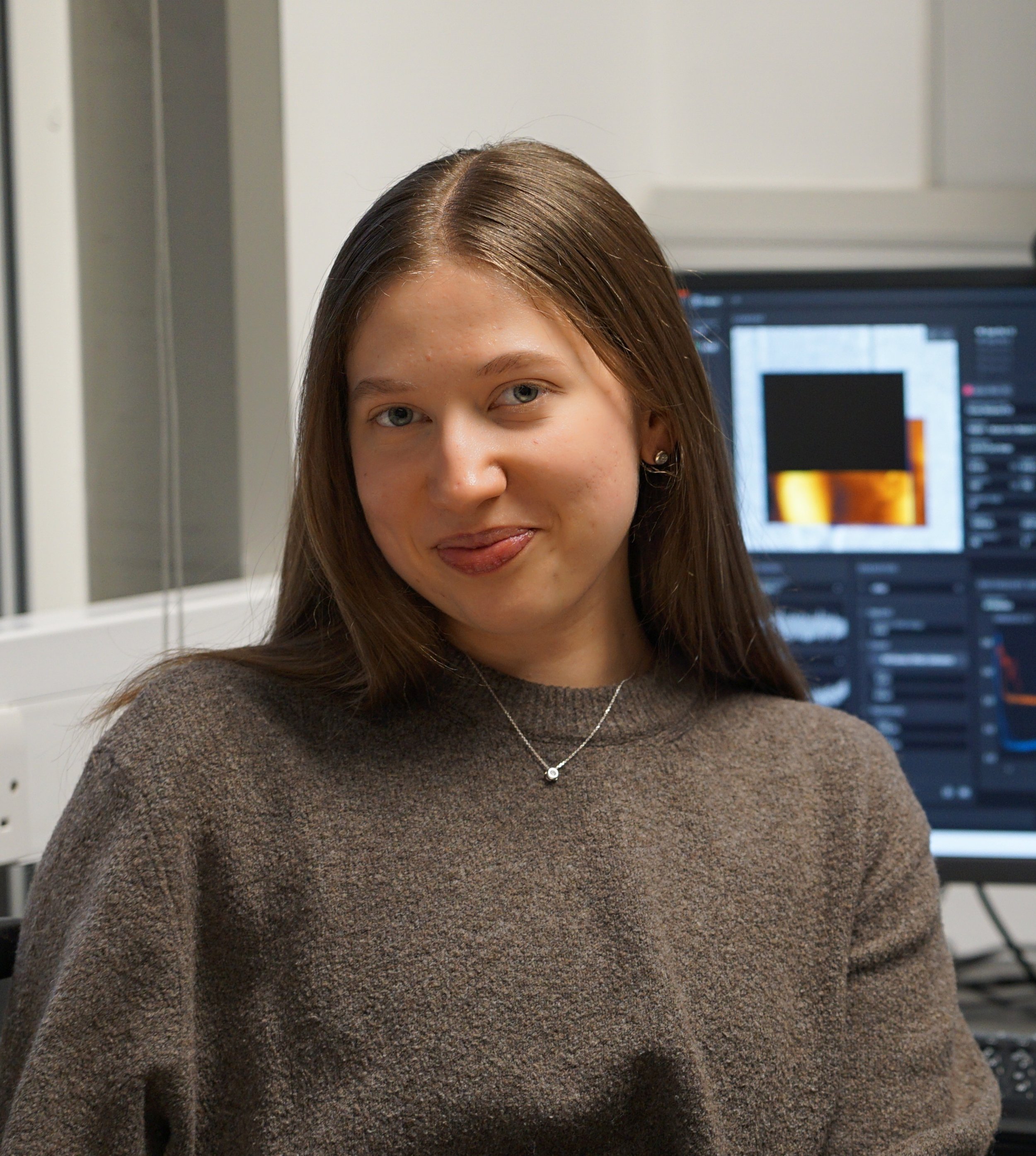
Dr Holly Linford

Holly Linford is a Postdoctoral Researcher in Dr Antonio Benedetto’s NanoBioPhysics lab at University College Dublin, Ireland, investigating the interactions of ionic liquids (ILs) with biomembranes and live cells by atomic force microscopy and other approaches, as part of a 4-years cross-disciplinary major research grant recently awarded to Dr Benedetto by Science Foundation Ireland. ILs are known to interact with the cell membrane and this is thought to be key to their toxicity. By using AFM in combination with biological assays, the group aims to determine the mechanism of action of ILs and identify effects at subtoxic concentrations.


Holly Linford


Recent AFM-related papers:

Chu J., Metcalfe P., Linford H. V., Zhao S., Goycoolea F. M., Chen S., Ye X., Holmes M. & Orfila C.(2022) Short-time acoustic and hydrodynamic cavitation improves dispersibility and functionality of pectin-rich biopolymers from citrus waste Journal of Cleaner Production 330, 129789

Rongkaumpan G., Amsbury S., Andablo-Reyes E., Linford H., Connell S., Knox J. P., Sarkar A., Benitez-Alfonso Y. & Orfila C. (2019) Cell Wall Polymer Composition and Spatial Distribution in Ripe Banana and Mango Fruit: Implications for Cell Adhesion and Texture Perception Frontiers in Plant Science 10

Biography: Holly began her journey in science in the chemistry department of Durham University (UK) and there discovered a love for advanced microscopy during her master's project. Following on from that, she took up AFM as part of her PhD at the University of Leeds (UK) with Dr Simon Connell. In this, Holly used AFM, along with a wide range of other biophysical and biochemical techniques, to probe the polymers and interactions that play a role in inter-cell adhesion in plant tissue. After completing her PhD, Holly moved to Dublin to join the group of Dr Antonio Benedetto at UCD, continuing to use AFM as a key technique for mechanical characterisation of cells on the nanoscale, this time looking at the interactions of ionic liquids with the mammalian cell membrane.

Twitter: @HollyVLinford

LinkedIn: https://www.linkedin.com/in/holly-linford-5bb280159/

Webpage: https://www.antoniobenedetto.eu/Members/


Are you a woman conducting AFM research or know of someone you would like to nominate to be featured in our next #WomenInAFM campaign? Contact us at community@nunano.com!

Dr Ana-Maria Zaske

Ana-Maria Zaske is the director of the Atomic force microscopy (AFM) core facility in the Internal Medicine Cardiology Division at the University of Texas Health Science Center at Houston, TX. USA. Her laboratory has emerged as a key platform for studying the morphological and nanomechanical properties of living biological systems.

The mission of Ana-Maria’s AFM core facility is to explore all meaningful ways to assess experimental trials to mitigate diseases and facilitate advanced life science research. She explains that the investigation of nanomechanical properties, at the single-cell and single-molecule level, is already advancing the development of new targeted diagnostics and therapies. She has the ability to image, probe and manipulate biological systems to address the needs of scientific and medical investigations.

An area of major interest in her lab is to evaluate the effects of several physiological processes by measuring the elastic response on cells and tissues. She mentioned that the elasticity of the cell membrane can vary between cell types as a function of growth, differentiation, disease or treatment. She is able to evaluate the effects of a drug in cancer cells, analyse the efficiency of skin healing, or simply observe the changes in DNA structures, all at the nano-metric scale.

Additionally, her AFM facility can map the distribution of elastic responses on the sample surface by combining force curve measurements with topographical imaging. She emphasises that the unique properties of Atomic Force Microscopy offer unlimited applications in medical research. That this technology can provide valuable insights about the mechanisms of a disease and assess a successful treatment pathway for its cure.


Biography: Ana-Maria received her PhD from the University of Manchester Institute of Science and Technology, in the UK where she was first trained in high resolution Atomic Force Microscopy. She went back to her native country and used Atomic Force Microscopy to characterize membrane receptors in follicular cells, as Research Associated in the Laboratory of Molecular and Cellular Neurobiology in the UNAM Mexico. In 2009 she was an award winner in recognition of her Scientific Excellence for presenting one of the 5 best posters at the “Seeing at the Nanoscale VII Conference” held in Santa Barbara CA by the VEECO company. She later joined UTHealth and became the director of the AFM core facility, as a part of the Internal Medicine Cardiology Division in Houston, TX. During the past 15 years, Dr. Zaske has gained relevant experience in the operation and applications of Atomic Force Microscopy to promote biomedical research.

LinkedIn: https://www.linkedin.com/in/ana-maria-zaske-phd-research-scientist-762aab52/   

Webpage: https://uthealth.corefacilities.org/service_center/show_external/3535?name=uthealth-atomic-force-microscopy

Debismita Dutta

Debismita Dutta is a 3rd year PhD student at the Advanced Materials and Surfaces Group, Tyndall National Institute, University College Cork, Ireland. She studies Aurivillius phase multiferroic thin films for novel data storage applications in her research project funded by Science Foundation Ireland. Magnetoelectric multiferroics are a type of material that showcase coupled ordered parameters of ferroelectricity and ferromagnetism. In essence, their magnetic polarization can be switched with an electric field, and vice versa, in a manner that retains the switched state even in the absence of the said field- hence becoming 'memory'. These materials also hold potential for devices with multiple memory states, which can store and process more than 8 times the amount of data in the same volume of thin film than current devices. Her work focuses on optimizing industry-relevant synthesis processes like DLI-CVD (Direct Liquid Injection-Chemical Vapor Deposition) to tackle the synthesis challenges that stand in the way of realizing a device roadmap for multiferroic devices. She uses Scanning Probe Microscopy techniques like Piezoresponse Force Microscopy (PFM), Variable Field PFM, Scanning Kelvin Probe Microscopy (SKPFM) and Atomic Force Microscopy (AFM) to study the grain, domain, and multiferroic characteristics of Aurivillius phase thin films. 


Debismita Dutta


Recent AFM-related paper:

Keeney L., Colfer L., Dutta D., Schmidt M. & Wei G. (2023) What lies beneath? Investigations of atomic force microscopy-based nano-machining to reveal sub-surface ferroelectric domain configurations in ultrathin films Microstructures 3:2023041.

Biography: Debismita is an Indian researcher who is passionate about exploring exciting new physics in materials by manoeuvring their chemical morphology. She holds a B.Tech in Chemical Engineering and has been very curious about materials research since her undergraduate days. Having done multiple competitive research internships in institutes like National Chemical Laboratory (NCL), as well as Korea Institute of Science and Technology (KIST), she knew she wanted to do a PhD. in materials science. Now she works in Micro Nano Systems in Tyndall National Institute under the supervision of Dr. Lynette Keeney and Dr. Michael Nolan in an SFI funded project. Besides her research, she also enjoys participating in public engagement events, some of which include Famelab Ireland, where she was a national finalist, and 3 Minute Thesis (3MT), where she was the university runner up. She enjoys talking about her work, and would love to discuss her research with you over email or on her social media handles listed below.

Email: debismita.dutta@tyndall.ie

Twitter: @Debbie7D

LinkedIn: https://www.linkedin.com/in/debismitadutta/


Are you a woman conducting AFM research or know of someone you would like to nominate to be featured in our next #WomenInAFM campaign? Contact us at community@nunano.com!

Professor Zoya Leonenko

Zoya Leonenko is a well-known expert in biophysics and scanning probe microscopy and is the author of over 90 scientific publications and several book chapters.

Zoya leads a nanoscale biophysics research group at the University of Waterloo, Canada. She uses advanced scanning probe microscopy and other biophysical methods, such atomic force microscopy (AFM) and Kelvin probe force microscopy (KPFM), nanoscale electrophysiology, plasmonic sensing with SPR, to study biophysics of lipids and lipid-protein interactions, interactions of nanoparticles with lipid membrane and monolayers, and to develop novel application of lipid films in biomedical nanotechnology. Her team provided significant advancement of knowledge in understanding the role of nanoscale structure of lipid membrane in molecular mechanisms of Alzheimer’s disease (AD). Recently together with collaborators she initiated a new research program in quantum biology, which has a focus to uncover new quantum phenomena in biology and neuroscience.


Zoya Leonenko


Recent AFM-related papers:

Biography: Zoya has PhD in Chemical Physics and is a Professor at University of Waterloo, Canada. She holds a joint position in the Department of Physics and Astronomy and the Department of Biology and is a member of the Waterloo Institute for Nanotechnology (WIN). Zoya joined the University of Waterloo in 2007. Prior to this, she worked at the University of Maryland at Baltimore, USA and the University of Calgary, Canada as an Assistant Professor. She was also a recipient of an Invited Professorship Award from the University of Burgundy, Dijon, France in 2012 and NSERC University Faculty Award in 2008-2012 and currently holds University Research Chair Award. Leonenko’s laboratory is supported by Natural Sciences and Engineering Research Council of Canada (NSERC), New Frontiers in Research Fund (NFRF) and Canada Foundation for Innovation (CFI) among other grants. She has made a significant impact on the establishment and subsequent rapid rise of the Biophysical Society of Canada (BSC) by organizing its very first annual meeting at Waterloo in 2015 and served as its President in 2019-2021.

LinkedIn: https://www.linkedin.com/in/zoya-leonenko-0b4444a4/

Webpage: https://uwaterloo.ca/leonenko-research-group/profile/zoya-leonenko